Anatomy of Biceps Femoris —
The biceps femoris (Latin: musculus biceps femoris) is a long and thick two-headed muscle of the lower limb located in the posterior compartment of the thigh.It is the most lateral muscle of the posterior compartment stretching between the hip bone, femur and fibula.Together with the semitendinosus and semimembranosus, the biceps femoris is also known as one of the hamstring muscles.

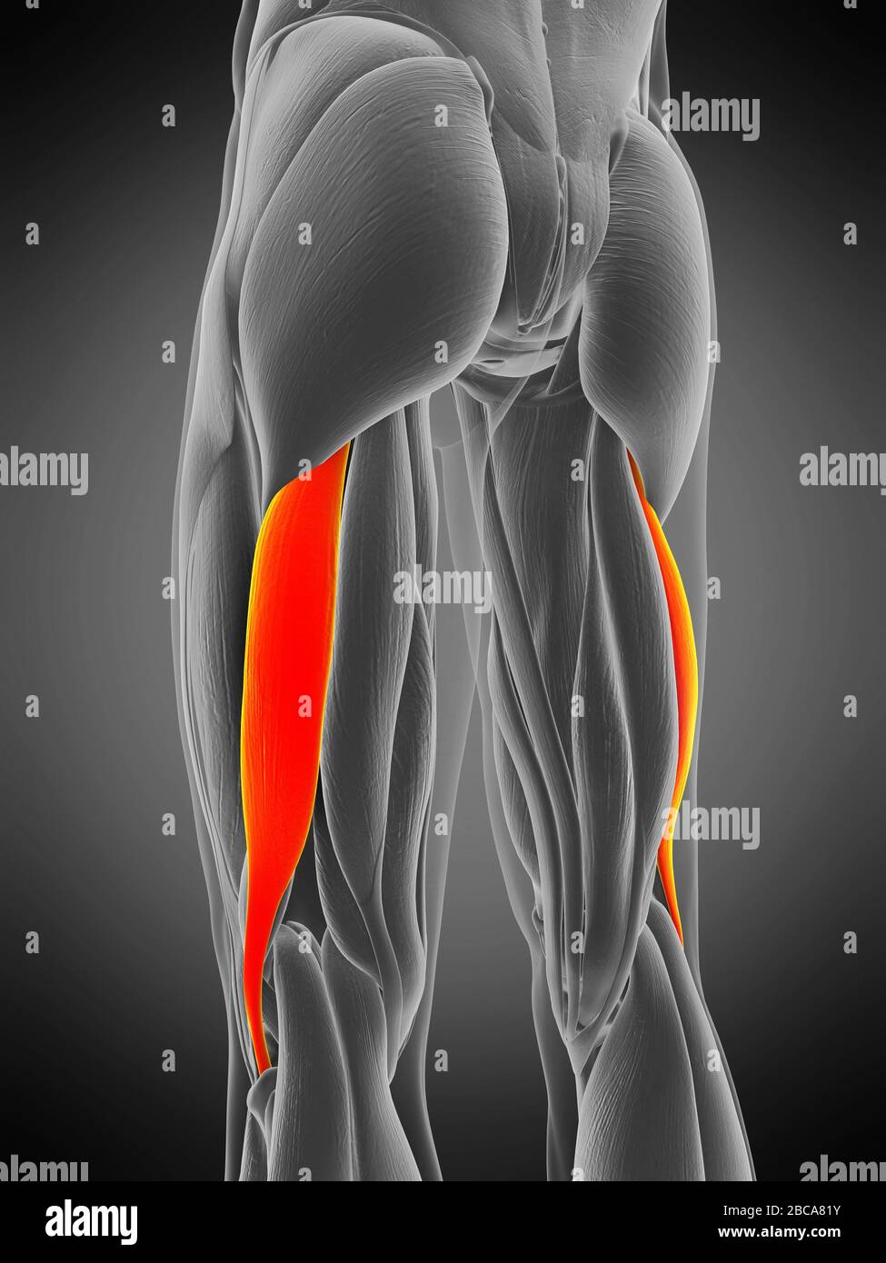
Biceps fémoral Photo Stock Alamy
The biceps femoris is a muscle within the posterior compartment of the thigh. It has two heads (long head and short head) and is the most lateral of the muscles in the posterior thigh. The common tendon of the two heads can be felt laterally within the popliteal fossa (posterior knee region). Attachments :
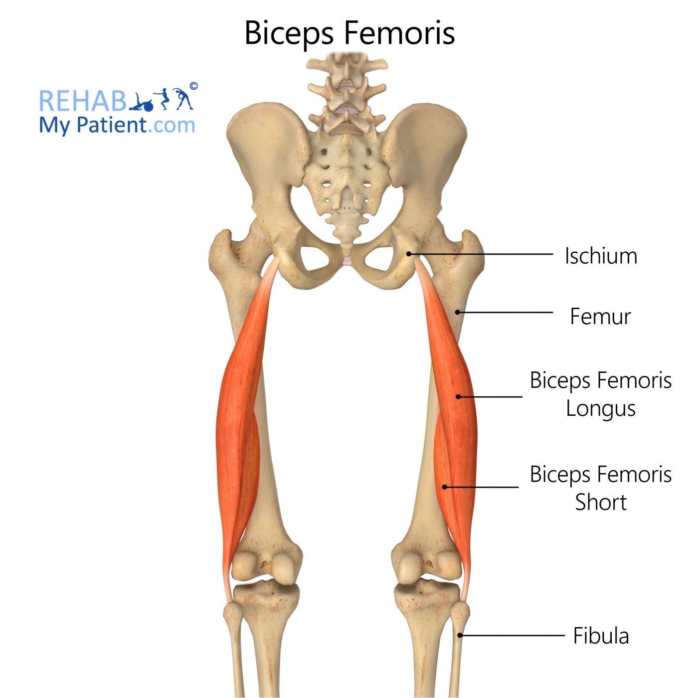
Biceps Femoris Rehab My Patient
The biceps femoris is one of the large muscles in the posterior compartment of the thigh and a component of the hamstrings. It has a long and a short head, each with different functions and innervation. Its medial border forms the superolateral border of the popliteal fossa. Summary origin long head: medial facet of the ischial tuberosity
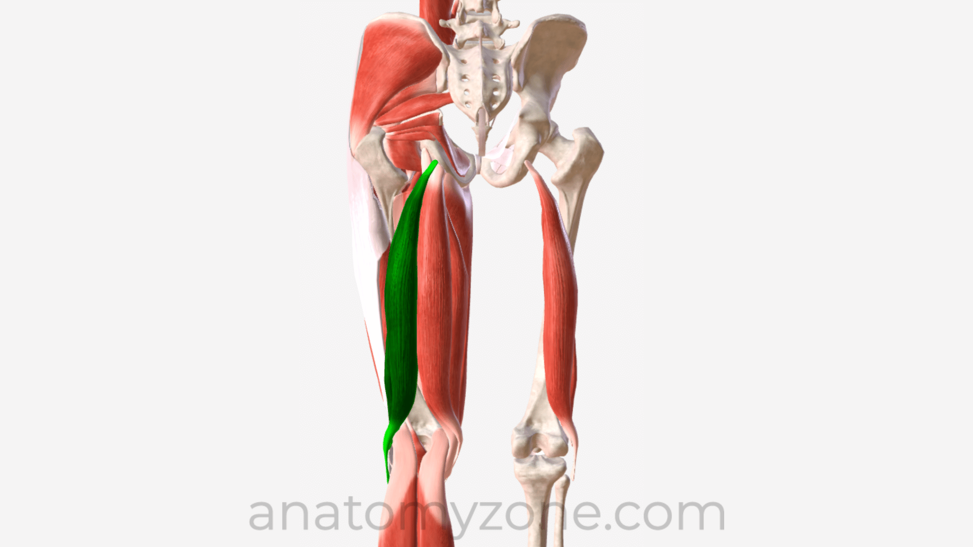
Biceps Femoris Origin, Insertion, Action, 3D Model AnatomyZone
Description Biceps femoris is a muscle of the posterior compartment of the thigh, and lies in the posterolateral aspect. It arises proximally by two 'heads', termed the 'long head' (superficial) and the 'short head' (deep). It is part of the hamstrings. [1] Anatomy Origin Long head: ischial tuberosity [3]
Musculus biceps femoris DocCheck
The biceps femoris is the most lateral of the muscles in the back of the thigh, i.e., it is the closest to the outer side of the leg. The muscle is innervated by two different divisions of the sciatic nerve: the common fibular division (which is also called the common peroneal nerve) and the tibial division.
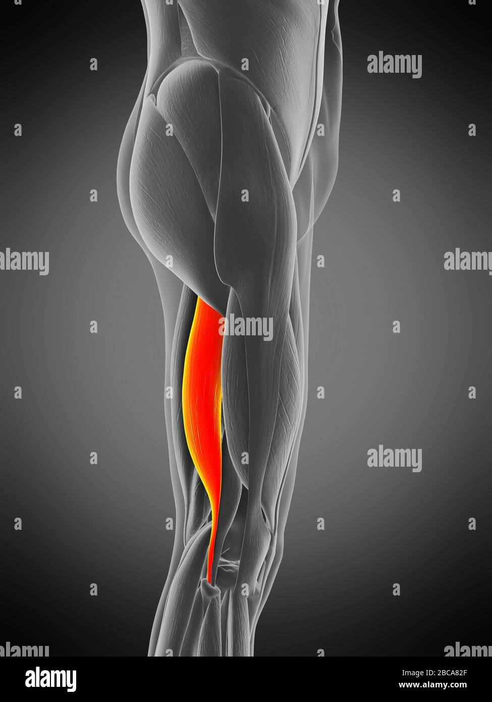
Biceps Femoris Longus Muscle Photograph by Sebastian Kaulitzki/science Photo Library Fine Art
Overall, the biceps femoris muscle contributes to the formation of the popliteal fossa, where the muscle and tendon form its superolateral boundary. The term "hamstrings" is the collective name given to the long head of biceps femoris, semitendinosus, and semimembranosus muscles. These three muscles share similar features, including:
Biceps femoris longus muscle, illustration Stock Photo Alamy
Der Musculus biceps femoris (zweiköpfiger Oberschenkelmuskel) ist ein zweigelenkiger Muskel an der Rückseite des Oberschenkels. Gemeinsam mit dem M. semimembranosus und dem M. semitendinosus bildet der die ischiocrurale Muskelgruppe, die sowohl auf das Kniegelenk als auch auf das Hüftgelenk wirkt. Inhalt Verlauf und Versorgung Funktion Klinik
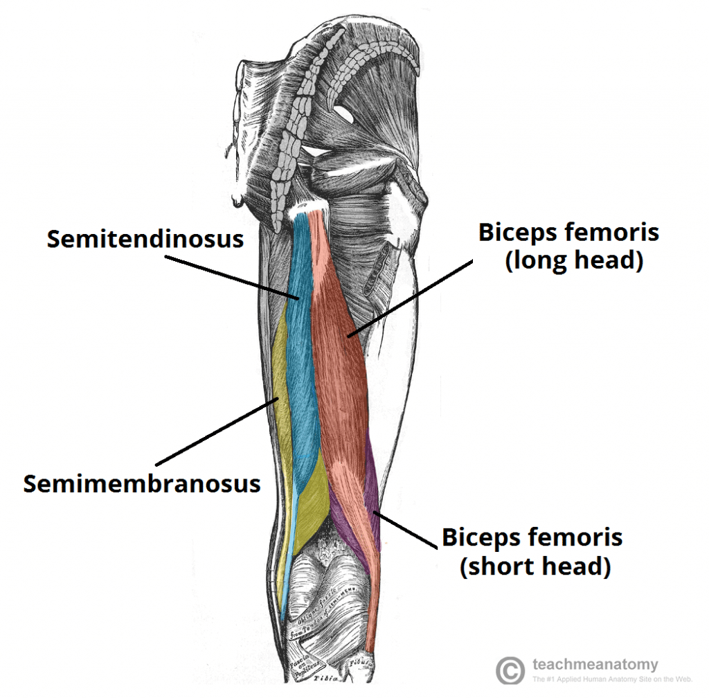
Biceps Femoris Attachments Actions TeachMeAnatomy
Objective: The aim of the present study was to compare the fascicle length, pennation angle, muscle thickness and stiffness of the biceps femoris long head, and eccentric hamstring strength between injured dominant limbs, injured non-dominant limbs, uninjured dominant limbs and uninjured non-dominant legs in previously injured players, and between dominant and non-dominant legs in uninjured.
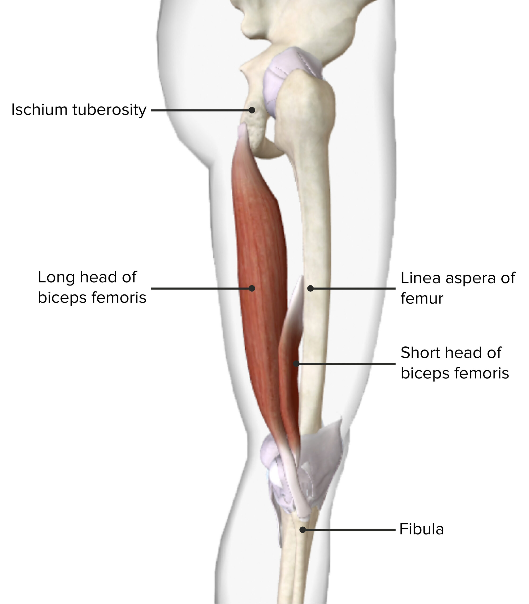
Thigh Anatomy Concise Medical Knowledge
The biceps femoris ( / ˈbaɪsɛps ˈfɛmərɪs /) is a muscle of the thigh located to the posterior, or back.

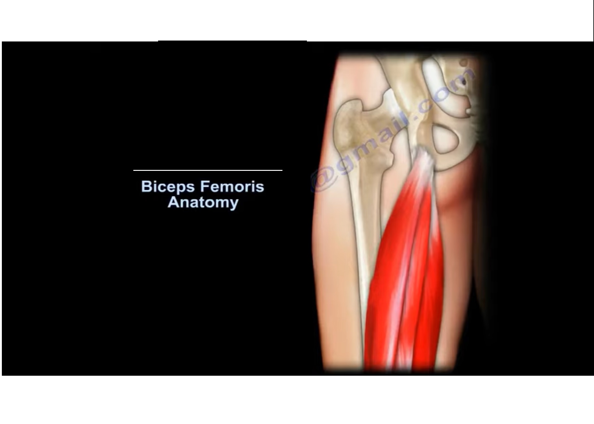
Biceps Femoris Anatomy Origin, Insertion & Action YouTube
⭐ Biceps Femoris Muscle ⭐💪 Origin (long head): Ischial tuberosity💪 Origin (short head): Linea aspera and the lateral supracondylar line of the femur💪 Inse.

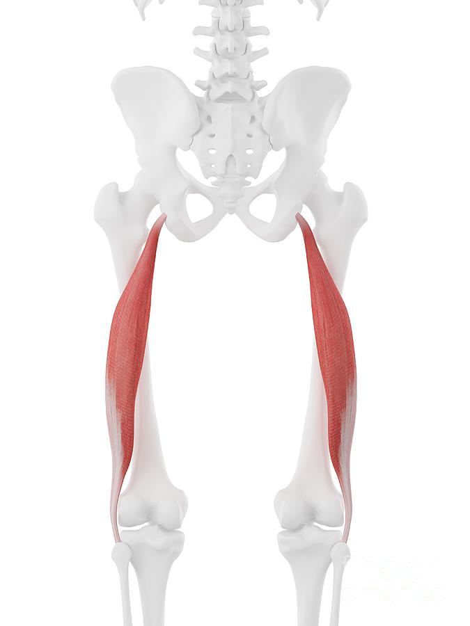
Biceps femoris longus muscle, illustration Stock Image F029/5137 Science Photo Library
The biceps femoris is a long muscle in the posterior compartment of the thigh responsible for movement at both the hip and knee joints. Along with the semitendinosus and semimembranosus, the biceps femoris makes up the hamstrings muscle. The muscles of the hamstring border the popliteal fossa, which is a triangular space behind the knee.

Muscle Breakdown Biceps Femoris
Key Features & Anatomical Relations Actions & Testing List of Clinical Correlates References Quick Facts Origin: Ischial tuberosity. Insertion: Head of fibula. Action: Flexes and laterally rotates leg at knee joint; extends thigh at hip joint. Innervation: Tibial division of sciatic nerve (L5-S2).

Anatomical model showing the biceps femoris muscles Stock Photo Alamy
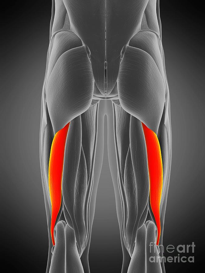
The biceps femoris is a double-headed muscle located on the back of thigh. It consists of two parts: the long head, attached to the ischium (the lower and back part of the hip bone), and the.
Biceps Femoris Longus Muscle Photograph by Sebastian Kaulitzki/science Photo Library Pixels
Dr. Ebraheim's educational animated video describes the condition of biceps femoris muscle anatomy.Follow me on twitter:https://twitter.com/#!/DrEbraheim_UTM.
Biceps femoris longus muscle, illustration Stock Photo Alamy
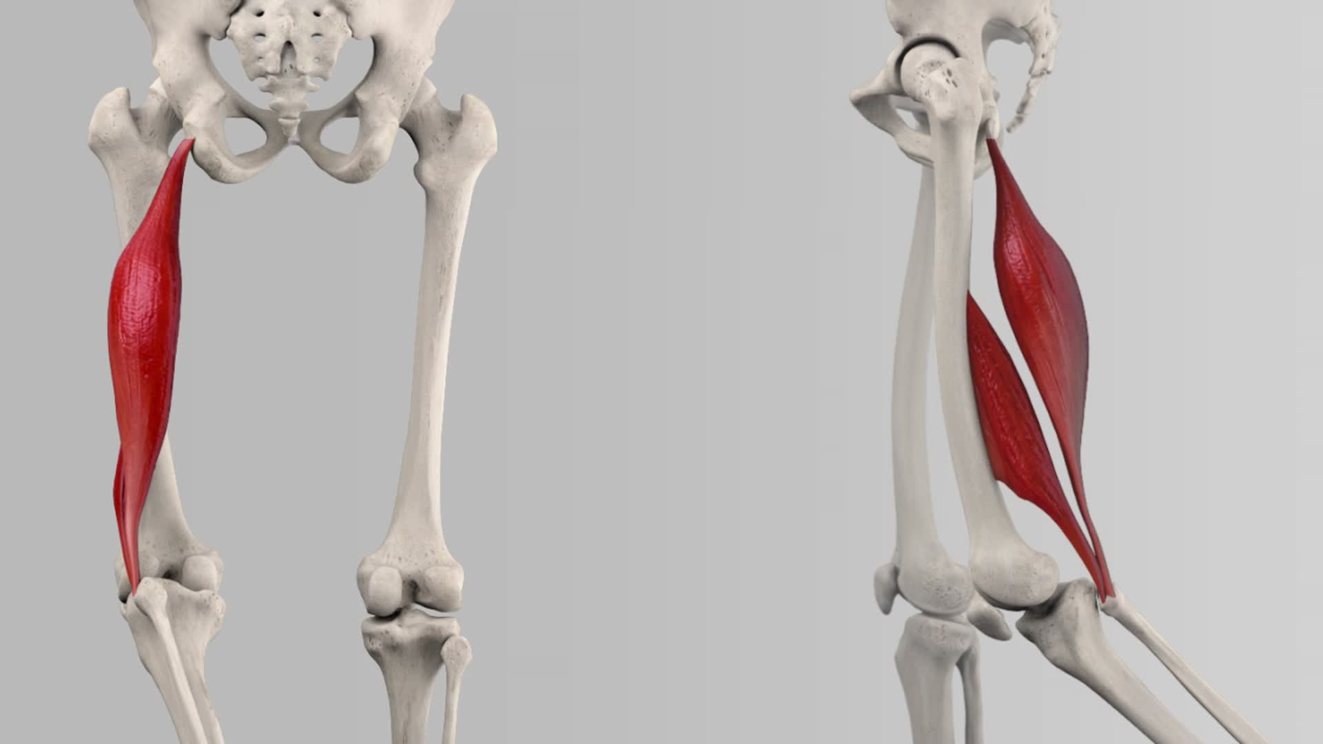
The biceps femoris tendon attaches the muscle bellies of the long and short heads of the biceps femoris muscle to the head of the fibula. This insertion site allows the biceps femoris muscle to be able to: - flex the leg at the knee joint; - laterally rotate the leg at the knee joint while this joint is held in a semiflexed position.
Biceps femoris Origin, insertion, innervation, function Kenhub
What is the Biceps Femoris? In more technical terms, the biceps femoris is a two-headed (hence "biceps") skeletal muscle, with one superficial head and the other being far deeper in the leg. The origin point is in the femur, while the distal attachment point lies in the fibula of the calves.